Tooth Histology Labeled Biology Diagrams Tooth anatomy (anterior view) The tooth anatomy is an interesting but challenging topic that demands the respect of any health science student or professional. The human teeth are quite special because they grow twice during a lifespan, are essential structures for the mechanical digestion of food, and support certain facial features.

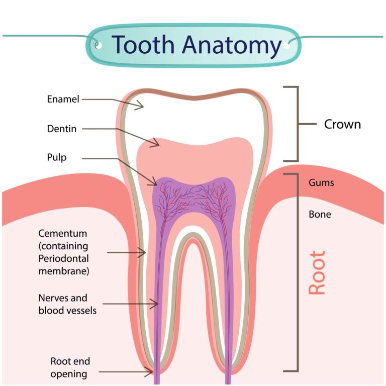
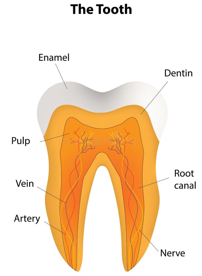
The crown refers to the part of a human tooth that is visible to us. The enamel, dentin, cementum, pulp, root, periodontal ligaments, etc., are important parts of the tooth structure. Bodytomy provides labeled human tooth diagrams to help you understand the human tooth anatomy.
Tooth anatomy: Names, types, structure, arteries, nerves Biology Diagrams
Atlas of dental anatomy: fully labeled illustrations of the teeth with dental terminology (orientation, surfaces, cusps, roots numbering systems) and detailed images of each permanent tooth These fully annotated anatomical illustrations are presented as a comprehensive atlas of the dental anatomy specifically designed for students in Tooth anatomy, labeled diagram. Tooth anatomy from outside and in cross section, labeled drawing. Dental checkup icon . Magnifying glass on a molar tooth, illustration. Plaque and tooth decay, medical drawing. Tooth decay stages diagram. Periodontal Disease, labeled diagram.
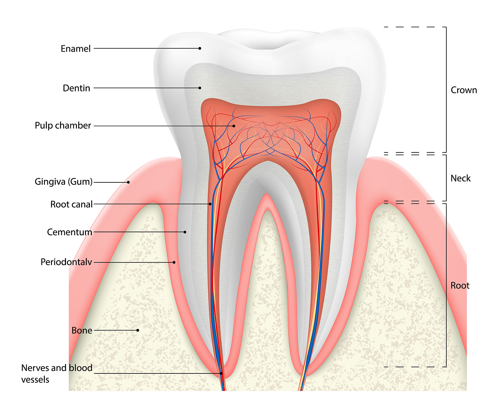
Information About the Human Tooth Anatomy With Labeled Diagrams. It is covered with enamel. The part of the tooth that cannot be seen and anchors the tooth to the bone is referred to as the root. It is covered by cementum. The area where the crown and the root meet is known as the cementoenamel junction (CEJ) or the neck/cervical line of the tooth. Tooth Anatomy Crown. In dentistry, the crown is the top part of a tooth covered by enamel, visible when you smile. Accidents or decay can cause it to chip or break. Dentists use artificial crowns to cover damaged teeth or implants. Bridges are used to fill the gaps when a tooth is missing. They can be attached to nearby natural teeth or implants.
Medical Information Illustrated Biology Diagrams
Print this Tooth Anatomy free using your home or office laser or inkjet printer and share with students at dental care class. Includes 1 labeled sheet to study tooth anatomy. Learn to identify tooth anatomy like the enamel, pulp cavity, dentin, gingiva, root canal, crown, root, periodontal membrane, cementum and many more.
